
Why on earth would I do a post about Osteosarcoma?
I’m posting up this information because I was on the receiving end of “your dog has Osteosarcoma” one of the “C” words you hope you never hear. Yes that is correct, my beloved Norman who will be 13 on November 3rd if he makes it that long has bone cancer. It all started in May when he was limping on his front right leg more than just his old age limping from time to time. I gave it some time to see if it was going to go away but after a couple of weeks it wasn’t going away. Being a technician and knowing too much about diseases I believe I was in denial and knew what was going on but if I choose to ignore it it will go away right? Wrong it doesn’t go away. I took Norman to work with me for a examination. He has some muscle wasting in the leg that he was limping on and was painful to the touch. Radiographs were in order.
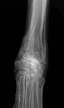
Here are the xray’s we took.

The lytic lesion is under the “bad Spot” text. The lower part of the bone going down is normal. This bone is the humerus.

This xray was taken with Norman on his back.
We pretty much knew after looking at the xrays that it was Osteosarcoma but we needed confirmation. In order to get a confirmation diagnosis a bone biopsy is in order. Monday June 3rd the orthopedic surgeon came and did his bone biopsy. For this he needed to be sedated so he wouldn’t feel it as a chunk of his bone would be removed using a big needle. While Norman was sleeping he was relaxed enough for a tumor in his abdomen to appear. That meant more xrays to find out what the tumor was attached to. The tumor is on his spleen. We took some needle aspirates to send to the pathologist to find out if the tumor is cancerous or not but the results were inclusive. When you are dealing with a organ that deals with blood you get blood and it is hard to tell what is what. So we don’t know if the tumor is malignant or benign. We do know that this tumor isn’t related to the Osteosarcoma as Osteosarcoma usually spreads to the lungs. So Norman has two diseases going on.
June 5th is when I got the dreaded news that no one wants to hear. I was devastated and it has taken me this long to tell Norman’s story. I’ve have had friends ask what my plan was with Norman and where do I go from here. It was suggested that I do a blog post on it so that is what I am doing.
Since Norman is 12.5 years old and had two young dogs at home to crash into him we have made a decision not to amputate his arm. You would amputate the arm to relieve the pain. You won’t be curing the cancer but taking away the painful bone. If you amputate you really should do chemotherapy as when you find the cancer in the bone more than likely it was already spread to the chest so you need to stop the development of the disease. Being a front leg and the leg where he bears most of his weight on we don’t want to amputate and have to have the risk of the young dogs hurting him. That is no way for him to life the rest of his life. The double wammy is that he still has that tumor on his spleen. We could do surgery on the spleen and remove it but what would be the point when he has cancer in another location in his body. If he had one or the other surgery on either one might be a option but since he has two things going on surgery is not a option for us. Norman is on pain relievers and still had a slight limp. When his limp gets worse we will make a decision at that time to say goodbye.
As many of you know Norman is my soulmate, the light of my life and by best friend. He was a gift from the day we brought him home from the breeders at 7 weeks of age that keeps on giving us unconditional love each and every day. He has been the best dog we could ever ask for and our gift to him is to not let him suffer. He has given so much to us we need to say goodbye to him before the times get tough. We owe him that much. He has had the best life with nearly any problems I don’t want his ending to be any different. I want to remember him in his young glorious years when he was full of life, hunting and running hunt tests and loving every minute. I don’t want to remember his last days on earth in pain with the look in his eyes that say you need to let me go. It is going to be the hardest thing I will ever have to do but I owe it to him.
We have been taking one day at a time. I kiss and hug him each day like it is his last. We do special things for him as we don’t know how long he will be around. He hasn’t a clue what is going on. He is a happy go lucky guy that still perks his ears up when we go out to train. He get’s a very short mark and the distance doesn’t matter to him. As long as he gets to retrieve a duck or a bumper he is in his glory.

I’m ready to go train!

I still got it!

Happiness is feathers in the mouth.

Happy boy!
The above pictures were taken after we got the results. Our friends from MN came down for the weekend and we did a little mock hunt test and gave Norman 2 live flyers. It was a chance for Mike to say goodbye to Norman. He has hunted with Norman in Canada and has one of his granddaughters. I hope we don’t have to say goodbye to Norman for many days or months but in the end I know that I have given him the best days of his life as he has done for me and he will never be forgotten.
Below information was taken from Veterinary Partner. They can explain Osteosarcoma better than I can.
Osteosarcoma (Canine)
What is Osteosarcoma?
Osteosarcoma is by far the most common bone tumor of the dog, usually striking the leg bones of larger breeds. Osteosarcoma usually occurs in middle aged or elderly dogs but can occur in a dog of any age; larger breeds tend to develop tumors at younger ages.
- Osteosarcoma can develop in any bone but the limbs account for 75-85% of affected bones. Osteosarcoma of the limbs is called appendicular osteosarcoma. It develops deep within the bone and becomes progressively more painful as it grows outward and the bone is destroyed from the inside out. The lameness goes from intermittent to constant over 1 to 3 months. Obvious swelling becomes evident as the tumor grows and normal bone is replaced by tumorous bone.
- Tumorous bone is not as strong as normal bone and can break with minor injury. This type of broken bone is called a pathologic fracture and may be the finding that confirms the diagnosis of bone tumor. Pathologic fractures will not heal and there is no point in putting on casts or attempting surgical stabilization.
How do we Know my Dog Really has an Osteosarcoma?
Radiographs (x-rays)
Compare bone consistency above the tumor and notice how the abnormal bone looks eaten away by the tumor.
One of the first steps in evaluating a persistent lameness is radiography (x-rays). Bone tumors are tender so it is usually clear what part of the limb should be radiographed. The osteosarcoma creates some characteristic findings.
- The lytic lesion – looks like an area of bone has been eaten away.
- The sunburst pattern – shows as a corona effect as the tumor grows outward and pushes the more normal outer bone up and away.
- A pathologic fracture may be seen through the abnormal bone.
- Osteosarcoma does not cross the joint space to affect other bones in the joint.
In most cases, radiography is all that is needed to make the diagnosis but sometimes there are ambiguities.
Biopsy
A tiny section of bone can be removed for laboratory analysis. This type of analysis is considered to be absolute proof of diagnosis. The procedure is associated with some pain and some oncologists suggest that biopsy is not needed if the radiographs show an obvious bone tumor. If there is any question about the lesion on the radiographs, a bone biopsy should provide clear results.
Sometimes a bone tumor is surrounded by an area of bone inflammation, so it may be difficult to get a diagnostic sample and several samples must be taken. These samples are too small to cause a pathologic fracture.
Amputation of the affected bone is recommended for any tumor involving bone. When the malignant structure has been removed, it is submitted for biopsy and the diagnosis confirmed at that time. Biopsy before amputation is felt to simply add a painful procedure and, if possible, is reserved for tissue already amputated.
What if it isn’t Really an Osteosarcoma?
The location and radiographic appearance of the osteosarcoma in the limb are quite classic but there are a few outside possibilities that should at least be mentioned. There are only a few other possible conditions that cause lytic lesions in bone: the chondrosarcoma, or the squamous cell carcinoma, or the synovial cell sarcoma.
Chondrosarcoma
Chondrosarcoma is a cartilage tumor, possibly not as malignant as the osteosarcoma. The chondrosarcoma generally occurs on flat bones such as ribs or skull bones and is not usually found in the limbs. Still, should a chondrosarcoma occur in the limb, treatment recommendations would include amputation of the affected bone and biopsy of the tissue after amputation would allow for adjustments in chemotherapy.
Squamous Cell Carcinoma
The squamous cell carcinoma is a tumor of the external coating of the bone (called the periosteum). This is a very destructive tumor locally but it tends to spread relatively slowly. Again, a bone suspected of malignant tumor should be amputated, and the tissue then analyzed and then treatment adjustments should be made thereafter. The squamous cell carcinoma tends not to arise in the same bone areas as the osteosarcoma; it tends to arise in the jaw bones or in the toe bones.
Synovial Cell Sarcoma
This is a tumor of the joint capsule lining. Its hallmark is that it affects both bones of the joint. The osteosarcoma, no matter how large or destructive it becomes, will never cross over to an adjacent bone.
Fungal Bone Infection
Immitis is a fungus native to the Lower Sonoran Life Zone of the Southwestern United States. It is the infectious agent of the disease called San Joaquin Valley Fever or just Valley Fever. (More scientifically, the condition is called coccidioidomycosis.) In most cases, infection is limited to a few calcified lymph nodes in the chest and possibly lung disease. In some rare cases, though, the fungus disseminated through the body and can cause a very proliferative bone infection. The bone infection of coccidioidomycosis is proliferative and lacks the lytic lesions that are so typical of the osteosarcoma.
The bottom line is that bone malignancy should be treated with amputation followed by additional treatment. What that treatment is depends on what the bone tumor is.
TREATMENT OF OSTEOSARCOMA INVOLVES TWO ASPECTS: TREATING THE PAIN AND FIGHTING THE CANCER’S SPREAD.
How do we Treat the Pain?
Keep in mind that dogs are usually euthanized due to the pain in the affected bone. Treating the pain successfully will allow a dog to live comfortably.
Amputation of the Limb
Removal of the affected limb resolves the pain in 100% of cases. Unfortunately, many people are reluctant to have this procedure performed due to misconceptions.
- While losing a leg is handicapping to a human, losing one leg out of four does not restrict a dog’s activity level. Running and playing are not inhibited by amputation once the surgical recovery period is over.
- While losing a limb is disfiguring to a human and has social ramifications, dogs are not self-conscious about their image. The dog will not feel disfigured by the surgery; it is the owner that will need to adjust to the dog’s new appearance.
- Median survival time for dogs who do not receive chemotherapy for osteosarcoma is 4 to 5 months from the time of diagnosis regardless of whether or not they have amputation. Do you want your dog’s last 4 to 5 months to be painful or comfortable?
Limb-sparing Surgery
Limb-sparing techniques developed for humans have been adapted for dogs. To spare the limb and thus avoid amputation, the tumorous bone is removed and either replaced by a bone graft from a bone bank or the remaining bone can be re-grown through a new technique called bone transport osteogenesis. The joint nearest the tumor is fused (i.e., fixed in one position and cannot be flexed or extended.)
- Limb sparing cannot be done if more than 50% of the bone is involved by tumor or if neighboring muscle is involved.
- Limb sparing does not work well for hind legs or tumors of arm bone.
- Limb sparing works best for tumors of the forearm bone.
- Complications can include: Bone infection, implant failure, tumor recurrence, and fracture.
Radiotherapy for Pain Control
Radiation doses can be applied to the tumor in three doses (the first two doses should be given 1 week apart, the second two doses 2 weeks apart.) Improved limb function is usually evident within the first 3 weeks and typically lasts 4 months (many oncologists report a range of 0 to 19 months.) When pain returns, radiation can be re-administered for further pain relief if deemed appropriate based on the stage of the cancer at that time.
- When pain is relieved in the tumorous limb, there is an increase in activity that can lead to a pathologic fracture of the bone.
- Radiotherapy does not produce a helpful response in about one-third of patients. (Remember, amputation controls pain in 100% of cases but if amputation is simply not an option, there is a two out of three chance that radiotherapy will control the pain.)
Drugs
At this time there are numerous analgesic medications available for dogs with this tumor. No single medication, however, is a match for the pain involved in what amounts to a slowly exploding bone. A combination of medications is needed to be reasonably palliative and should be considered only as a last resort if amputation or radiation therapy will not be pursued. There are several types of drugs that can be combined.
Non-Steroidal Anti-Inflammatory Drugs (NSAIDs)
These are anti-inflammatory pain relievers developed for dogs: carprofen, etodolac, deracoxib, meloxicam, firocoxib, and tepoxalin. These are typically given once or twice daily in tablet form at home. The patient should have good liver and kidney function in order to take medications of this class.
Bisphosphonates
This class of drug has become the standard of care in humans with bone tumors yet bisphosphonates have not become a common part of veterinary practice for this condition. Bisphosphonates act by inhibiting bone destruction, which in turn helps control the pain and bone damage caused by the bone tumor. The most common bisphosphonate in use for dogs is pamidronate, which is given as an IV drip over two hours in the hospital every 3 to 4 weeks. In humans, an assortment of potential side effects have emerged (fever, muscle pain, nausea all lasting 1 to 2 days in up to 25% of patients, renal disease in certain situations, low blood calcium levels, jaw bone cell death); these issues so far have not panned out as problems for dogs and cats. Because bisphosphonate seem to be well tolerated, relatively inexpensive, and useful in numerous bone-destroying cancers, we expect to see this class of drug used more and more in small animal practice.
Narcotic Pain Relievers
While these drugs do not have anti-inflammatory properties, they are well-known analgesics and have been used in an assortment of forms for thousands of years. They are particularly useful in chronic pain because they do not interact negatively with other pain relievers. Drowsiness is a potential side effect. Tramadolhas been particularly popular as part of a drug combination for bone cancer pain but there are other narcotics that might also be considered.
Miscellaneous Supplemental Pain Relievers
There are two drugs that have surfaced as additional pain relievers for animals with chronic pain: gabapentin and amantadine. Gabapentinworks on neurologic pain and is rapidly surfacing in the treatment of arthritis, surgical pain, and other chronic pain states. Amantadineworks by reducing what is called wind up, a phenomenon where nerves become sensitized to pain leading to the experience of pain from stimuli that normally do not cause pain.
These different drugs are often given together to create meaningful pain relief to the osteosarcoma patient when amputation and radiotherapy are not going to happen.
How do we Treat the Cancer?
Osteosarcoma is unfortunately a fast-spreading tumor. By the time the tumor is found in the limb, it is considered to have already spread. Osteosarcoma spreads to the lung in a malignant process called metastasis. Prognosis is substantially worse if the tumor spread is actually visible on chest radiographs, so if chemotherapy is being considered, it is important to have chest radiographs taken.
- Chemotherapy is the only meaningful way to alter the course of this cancer.
- Young dogs with osteosarcoma tend to have shorter survival times and more aggressive disease than older dogs with osteosarcoma.
- Elevations of alkaline phosphatase, one of the enzymes screened on a basic blood panel, bode poorly. These dogs have approximately 50% of the survival times quoted below for each protocol.
- The presence of tumor in lymph nodes local to the leg being amputated also bodes poorly. In the study by Hillers et. al published in the April 15th, 2005 issue of the Journal of the AVMA, median survival was significantly longer (318 days vs. 59 days) in dogs where the tumor was not evident in local lymph nodes at the time of amputation.
Cisplatin (given IV every 3 to 4 weeks for 3 treatments)
- The median survival time with this therapy is 400 days.
- Survival at 1 year: 30% to 60%
- Survival at 2 years: 7% to 21%
- Giving less than 3 doses does not increase survival time (i.e., if one can only afford one or two treatments, it is not worth the expense of therapy)
- Cisplatin can be toxic to the kidneys and should not be used in animals with pre-existing kidney disease.
Carboplatin (given by IV every 3 to 4 weeks for 4 treatments)
- Similar statistics to cisplatin but carboplatin is not toxic to the kidneys and can be used if the patient has pre-existing kidney disease.
- Carboplatin is substantially more expensive than cisplatin.
Doxorubicin (given IV every 2 weeks for 5 treatments)
- The median survival time is 365 days.
- 10% still alive at 2 years.
- Toxic to the heart. An ultrasound examination is needed prior to using this drug as it should not be given to patients with reduced heart contracting ability.
Doxorubicin and Cisplatin in Combination (both given IV together every 3 weeks for four treatments)
- 48% survival at 1 year
- 30% survival at 2 years
- 16% survival at 3 years.
What Exactly is “Median” Survival Time?
When a population is evaluated statistically, there are a number of ways the central tendency of the group can be evaluated. The median is the value at which 50% of the group falls above and 50% of the group falls below. This is a little different from the average of the group, though more people are familiar with this term. When you evaluate median survival times, you are looking at a 50% chance of surviving longer than the median (and a 50% chance of surviving less than the median).
What Does Chemotherapy Put my Dog Through?
Most people have an image of the chemotherapy patient either through experience or the media and this image typically includes lots of weakness, nausea, and hair loss. In fact, the animal experience in chemotherapy is not nearly as dramatic. After the pet has a treatment, you should expect 1 to 2 days of lethargy and nausea. This is often substantially palliated with medications like Zofran® (a strong antinausea drug commonly used in chemotherapy patients). These side effects are worse if a combination of drugs is used but the pet is typically back to normal by the third day after treatment.
Effectively, you are trading 8 days of sickness for 6 to 12 months of quality life. Hair loss is not a feature of animal chemotherapy.
Axial Osteosarcoma
While osteosarcoma of the limbs is the classical form of this disease, osteosarcoma can develop anywhere there is bone. Axial osteosarcoma is the term for osteosarcoma originating in bones other than limb bones, with the most commonly affected bones being the jaws (both lower and upper). Victims of the axial form of osteosarcoma tend to be smaller, middle-aged, and females outnumber males two to one.
In the axial skeleton the tumor does not grow rapidly as do the appendicular tumors, thus leading to a more insidious course of disease. The tumor may be there for as long as two years before it is formally diagnosed. An exception is osteosarcoma of the rib, which tends to be more aggressive than other axial osteosarcomas.
Treatment for axial osteosarcoma is similar to that for the appendicular form: surgery followed by chemotherapy. There is one exception, that being osteosarcoma of the lower jaw. Because of the slower growth of the axial tumor and the ability to remove part or all of the jaw bone with little loss of function or cosmetic disfigurement, it has been reported that 71% of cases survived one year or longer with no chemotherapy at all.
Additional information can be found at Bone Cancer Dogs, Inc., a nonprofit corporation.
Treating osteosarcoma is an area that not all veterinarians are comfortable performing. Discuss with your veterinarian whether referral to a specialist would be best for you and your pet.
Date Published: 3/29/2002
Date Reviewed/Revised: 08/12/2011